Menu
Videos of Various Genera Under The Microscope
Basic Microscope Recommendations and Magnification
Finding, Raising and Tending to Protozoa
Creating wet mount slides
Tips and Tricks for the Microscope
Recommended Books for Study
Camera and Video
Kahl, 1932 - 320 Drawings of Hypotrichida
Sorted to Genus by Stephen Cunningham, Baltimore, MD, USA
PDF document, 20 pages.
Acknowledgments
Help with Identification
David Seamer, Australia
David Seamer has written a series of identification quides to microscopic life found world-wide. Here is his website: http://davidseamer.com/
Bill Porter, Kentucky
Mr. Porter has plenty of informative protozoan videos on his YouTube channel:
https://www.youtube.com/user/BillPorter1456/videos
Bruce Taylor, Quebec
Mr. Taylor has a series of protozoa identification videos on his YouTube channel:
https://www.youtube.com/user/BruceDSTaylor/videos
Carlos Uruguay, Uruguay
Carlos has plenty of identification videos on his YouTube channel:
https://www.youtube.com/user/microuruguay/videos
Species Identification
Identifying protozoa and invertebrates can be difficult due to the countless varieties within the orders, families, and genera. Species identification takes into account many factors including type of microscope, DNA, RNA, number of kineties, etc., and is beyond the scope of this website.
In many cases, the examples on this page have been identified to genera with the extension of "sp." meaning unspecified species. In other cases, it was relativly easy to identify to species useing light microscopy.
|
| |
THROUGH THE MICROSCOPE
Microscopic Organisms
Click on a Photo to Watch Video
--------- Stop videos as needed ---------
Back to Menu |
 |
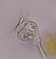
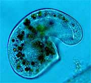
Limnias novemceras P. Meksuwan 2018
Rotifera, Flosculariidae.
Three adults were found at Patterson Park Lake, Baltimore, MD, USA. No larvae were found. Sizes ranged from 210um to 450um from the base of the foot to the tip of the corona.
The most distinguishing features of L. novemceras are the crescent-shaped lobes of the corona that normally don't extend very far from the tube opening giving the corona a somewhat flattened appearance, and the very wide dorsal gap.
Other features are a ringed transparent tube except at the base where is smooth and transparent, two long antennae that extend beyond the corona, and a dorsal plate with nine projections.
The dorsal plate is a stiff structure starting just beneath the ventral sinus (area between the crescent-shaped lobes of the corona) extending over the mastax (food grasping and crushing mechanism). The number of projections is a criteria to identifying all Limnias species. Other keys are tube structure and antennae length.
L. novemceras was first described in 2018 "Two new species of genus Limnias from Thailand, with keys to congeners (Rotifera, Gnesiotrocha)" by Phuripong Meksuwan, Rapeepan Jaturapruek and Supiyanit Maiphae. At that time the rotifer was only known from Thailand. |
 |
Limnias melicerta Weisse, 1848
Rotifera, Flosculariidae.
18 adults and 3 larvae studied were collected from various ponds around Baltimore, MD, USA. Length ranges from 150um (young tube) to 1000um measuring from the base of the foot to the tip of the corona. The worm-like larvae measured from 135um to 150um in length..
Diagnosis of Limnias species involves just three criteria: number of projections on dorsal plate; tube structure and ventral antenna length.
L melicerta lives in a transparent ringed tube (the base of which is without rings) that is attached to aquatic plants. The dorsal plate has seven projections and the antennae are short. L. shiawasseensis also has seven projections on the dorsal plate but the tube is not ringed and the antennae are long. A lobed corona with a deep ventral sinus brings food to the mouth.
The dorsal plate is a stiff plate starting at the deep ventral sinus (the area between the lobes of the corona) and extends over the mastax (the food grasping and crushing mechanism that pumps food into the esophagus). The mastax has three predominant teeth on each side of the trophi.
The foot is attached to the bottom of the tube by a pedestal which can be seen when fully extended. When startled, the foot contracts in accordion fashion bringing the corona well within the tube.
In females, eggs can be seen next to the foot inside the tube where they hatch into worm-like larvae divided into three regions: head, trunk and foot. Larvae leave the tube and swim around, using the cilia of the head and short cilia of the foot, looking for a place to attach. They have two distinct eye spots which disappear after the larvae settles. |
 |
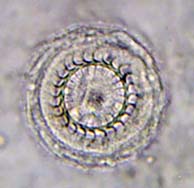
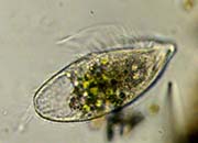
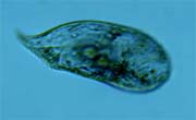
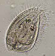
Glaucoma scintillans Ehrenberg, 1830
Ciliophora, Hymenostomatia, Tetrahymenida, Glaucomidae
Sixteen specimens of Glaucoma scintillans were found in a birdbath in Baltimore, Maryland, USA, that ranged in length from 50μm to 73μm.
The body shape is ellipsoidal and, in two examples, there are 38 ciliary rows. The oral opening features a row of cilia that curves around the anterior oral opening to the opposite side (species specific).
The macronucleus is large and centrally located. The contractile vacuole is in the lower third of the cell. Many round food vacuoles are present. Feeds on bacteria. |
 |
Loxodes striatus Englemann, 1862
Ciliophora, Karyorelictea.
26 individuals were found in various lakes, ponds and swamps around Baltimore, MD USA, ranging in size from 154μm to 255μm. Body shape is long, flat and flexible, with beak-like anterior rostrum.
The oral opening consists of mouth and pharyngeal basket of trichites.
There is an anterior and posterior macronucleus. There is no contractile vacuole.
Pigment granules, concentrated around the mouth and pharyngeal tube, are present and used for defense. The granules are extrusomes containing a toxic chemical.
Interesting facts about Loxodes:
- The macronuclei do not divide. Instead the micronuclei do all the work.
- There is no contractile vacuole in the Loxodes genera.
- Loxodes contains Müller vesicles - fluid filled vesicles containing a spherical center mass of barium salts, thought to be gravity sensors.
- Loxodes is the only fresh water genera in the Class Karyorelictea.
- Loxodes is one of the few ciliates that, in low oxygen or anoxic environments, are able to use nitrogen instead of oxygen as an electron acceptor for respiration.
- Juveniles have few Müller vesicles. As they grow, more vesicles develop.
- Loxodes striatus is the only binucleated species in the genus Loxodes where the two macronuclei are separated into anterior and posterior locations.
|
 |
Frontonia leucas (Ehrenberg, 1833)
Ciliophora, Peniculita.
Syn; Bursaria leucas; Frontonia vernalis; Ophryoglena magna; Ophryoglena vorax; Plagiopyla hatchi.
84 individual cells were collected from various ponds around Baltimore, Maryland, USA, including a bird bath. Length of the cells ranged from 150μm to 730μm.
Terms for study of ciliature are often used in reference to silver stained fixed cells. A kinetosome (basal body) is the protein structure at the base of each cilium that is dark when silver stained clearly showing ciliary patterns. A kinety is a row of closely connected cilia such as the longitudinal ciliary rows of ciliates. Polykineties are closely arranged brush-like cilia. The oral peniculi are composed of polykineties.
Frontonia leucas is a common ciliate found in lakes, ponds and streams. The dorso-ventrally compressed body is flexible, elongated ellipsoidal in outline, rounded at the poles, with the anterior end often slightly broader than the posterior. There are three peniculi (brush-like ciliated bands) on the right side of the mouth opposite the vestibular kineties shown in the video. The vestibular kineties are sometimes confused with the peniculi. A postoral groove of kineties stretches from the base of the mouth to the posterior end of the cell. A paroral membrane (undulating membrane) is a group of fused cilia into a moveable membrane-like sheet used to sweep food toward the cytostome (mouth) and extends from the very top anterior of the mouth to the base of the vestibular kineties.
The cell has longitudinal rows of cilia (kineties). The body is covered with many prominent undischarged trichocysts, each containing a protrusible filament, that reside just beneath the cell membrane and are used for defense and stunning prey.
The macronucleus is ellipsoidal and can be found either at cell center, in the posterior portion of the cell or slightly forward of the midline. One contractile vacuole is found about mid body on the margin of the cell and is often fed by radiating water canals.
Eats filamentous cyanobacteria, testate amoebas, rotifers, diatoms, desmids, flagellates and other ciliates. |
 |
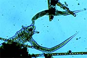
Zosterodasys transversus (Kahl, 1928) Foissner et al., 1994
Syn: Chilodontopsis (Chilodon); Chilodontopsis transversa; Chilodontopsis vorax, and several others as in Vďačný, Peter, Tirjaková, Eva (2012), "Taxonomic Revision and Neotypification of Zosterodasys transversus."
Two populations of Z. transversus were collected: Population I from a small stream at Howard County Fairgrounds, Howard County, MD USA (19 individuals) and Population II from Lost Lake, Patapsco State Park, Catonsville, MD, USA (2 individuals). The length of the individuals from both populations measured between 120 μm and 240 μm.
The anterior end of Zosterodasys is arched. The flexible cell tapers to a rounded posterior end. The ciliature runs lengthwise in evenly distributed rows throughout the surface and is interrupted by a band of longer cilia, known as the synhymenium or hypostomial frange, which runs transversely across the anterior part of the cell. The overall shape of the cell may vary.
The most striking feature of Z. transversus is the large circular cyrtos composed a mouth opening (cytostome) surrounded by nematodesmal rods arranged in a circle that penetrate deep into the cytoplasm.
The macronucleus in Population I is ellipsoidal and very close to the cyrtos whereas those of population II are more rounded and located mid-body. The macronucleus may vary in shape and location in various populations.
Several contractile vacuoles are distributed throughout the cell. Often a prominent posterior contractile vacuole is distinctly enlarged.
Z. transversus feeds on diatoms. |

|
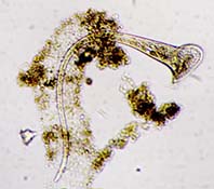
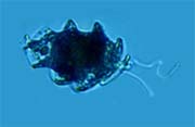
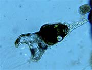
A species of the family Manuelophryidae (Dovgal, 2002)
Ciliophora, Suctoria.
A parasite of sessile peritrichs.
This rare species was found at Lost Lake, Patapsco State Park, Catonsville, MD, USA. Total body length ~60 um. The suctorian is feeding on the cytoplasm of Pseudovorticella chlamydophora.
Manuelophryidae are a family of ectoparasitic suctorians that prey on sessile peritrichs. The three genera of the family are Manuelophyra, Mistarcon and Pseudogemmides. There are few drawings representing the family save for Jankovsky 1997 " MISTARCON PARASITICUS - Ectoparasite Peritricha LAKE BAIKAL AND STATUS OF THE GENUS MANUELOPHRYA," Jankovsky 1997; and "EVOLUTION, PHYLOGENY AND CLASSIFICATION OF SUCTOREA (CILIOPHORA)" Igor V. Dovga 2002.
The example in the video does not resemble any of the species depicted in either publication, but the means by which the parasite penetrates the peritrich is the same: A single rod-like tentacle, about 7 μm in length, penetrates the host and feeds on the cytoplasm.
The longitudinally ciliated body is wider posteriorly, has one posterior contractile vacuole and a centrally located spherical macronucleus. There is no lorica. It wraps around and clings to the stalk of the host by means of cilia. |
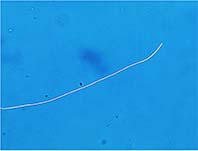 |
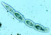
Actinomycetales (Buchanan, 1917)
Prokaryote, Actinobacteria.
Filamentous bacteria.
Collected from various lakes and streams around Baltimore, MD, these examples of filamentous bacteria were found in the anaerobic environment of bottom sediments.
Filamentous bacteria grow end to end like a chain, with each cell separated by a septum. Eventually the cells become a long filament or mycelium. The example in the video shows that the filaments are capable of worm-like movement which enables numerous strands to become entangled to form a colony. The individual chains wiggle around as though trying to gather more debris.
Flocculation is the process by which tangled colonies trap debris to form clumps or floc, which eventually drops to the bottom of a body of water. Waste water treatment facilities use flocculation to settle out debris in settlement tanks.
The numerous species of filamentous bacteria come in many shapes and sizes such as threads, string of beads, branching filaments, square filaments, curved and many more. |
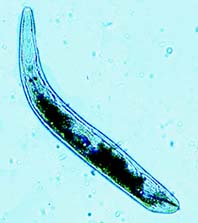 |
Gyratrix hermaphroditus Ehrenberg, 1831 - A Closer Look
Platyhelminthes, Polycystididae.
Flatworm
Collected from Patterson Park Lake, Baltimore, MD, USA, these common flatworms measured from 400 μm to 800 μm in length. Five specimens were examined with a microscope.
G. hermaphroditus is an oblong, highly flexible and contractile flatworm that when startled can retract into a ball. When swimming they can extend to 2 mm in length.
The ciliated epidermis is comprised of three cell layers: ectoderm (external), mesoderm and endoderm (internal). The muscular system originates from the mesoderm.
The two eyes are black, close together and prominent. In front of the eyes is a proboscis sheath that feels for food through the substrate. The proboscis sheath contains a sensitive hollow proboscis nerve cone, of nerves that can be withdrawn from the front of the proboscis sheath by long retractor muscles attached in front of the eyes.
The brain is just behind the eyes. An anterior nerve bundle is seen connecting the proboscis nerve cone to the brain, as well as lateral nerve cords.
The pharynx is behind the eyes at about the middle of the body. It appears as a large circle of strong muscle fibers. One video shows the worm sucking out the insides of a crustacean larva (nauplius). There is no anus. Waste goes out through the pharynx.
The circulatory system has no central beating heart. Instead, a clear nutritive liquid is circulated by means of body contraction.
At the tail end (posterior) of the worm a prominent penile stylet can be seen that is used for reproduction and defense. It simultaneously injects sperm, penis and stinger at the same time. It is connected to a poison gland that immobilizes prey.
A single greenish-brown teardrop-shaped egg is laid by the female. The egg is connected to the substrate by means of a stalk. The young worm vacates the egg case though a hatch at the top which closes when the egg case is empty. |
 |
Stentor roeselii (Ehrenberg, 1835) and the traveling AZM.
Ciliophora, Heterotrichida.
Synonyms: S. fimbriatus, S. fuscus, S. gracilis, S. magnus, S. roeseli f. stagnalis, S. viridis.
Collected from Patterson Park Lake, Baltimore, MD, USA, the length of the individuals found ranged from 400 μm to 1400 μm when fully extended and feeding. 44 individuals were studied. Most of the individuals were surrounded by a mucilaginous lorica composed of loose debris and waste.
Common and highly contractible, S. roeselii can vary in shape from oval swimming cells, to the fully extended, trumpet-like feeding cells. Sensory bristles are present along with the shorter body cilia throughout the length of the cell. It is a transparent colorless species with no pigment in the cortex. Alternating rows of pigment granules and clear stripes extend throughout the body length. There is no pigment within the granules in S. roeselii. There are no algae symbionts. When feeding, the cell attaches to debris or other structures by means of a holdfast.
Stentor eats by way of a membranellar band of cilia (adoral zone of membranelles = AZM) encircling the apical end spiraling around into a tightly spiraled oral opening leading to the gullet. The AZM encircles the ciliary lines of the peristomial bottom.
The macronucleus is vermiform (rope-like) and can extend well into the cell's posterior. While swimming, the macronucleus gathers in random folds depending on the length of the cell. A single contractile vacuole is located adjacent to the oral opening.
Early stages of sexual division are shown as well as late stage transverse division. During early division, a line of oral cilia (primordium) can be seen extending longitudinally from below the parent's oral spiral. The end of this primordium will form a secondary oral spiral representing a new daughter cell. In one example an additional contractile vacuole of the daughter cell can be seen. Eventually transverse division will occur with what looks like one Stentorcoming out of the oral end of another. |
 |
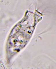
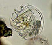
Orbopyxidiella susannae Schödel, 2004.
Ciliophora, Operculariidae
Ostracods, host to numerous sessile epizoic peritrich species, were collected from March through May from a thin layer of organic muck at the bottom of Patterson Park Lake, Baltimore, MD in about three feet of water. Hundreds of Ostracods were individually isolated and examined with a microscope.
Orbopyxidiella susannae is a solitary epizoic species that prefers to attach to the Ostracod Cypria ophthalmica. Hundreds of zooids were observed attached to the Ostracods.
Zooids ranged in length between 20 μm and 40 μm. The body is barrel shaped, elongated to slightly bloated and the pellicle has faint transverse striations. The body sits upon a very short non-contractile stalk anchored to the host by means of a small circular disc.
The peristome has no lip. A small cone-shaped peristomial disc rises slightly above and often leans to one side of the peristome. The infundibulum is deep at the longitudinal center of the cell. One contractile vacuole is located just above and near the infundibulum and a large spherical to ovate macronucleus is located near or above the center of cell.
When contracted, the peristomial disc withdraws into the cell, the peristome closes and the zooid leans over toward the surface of its host. |
 |
Epistylis anastatica Linnaeus, 1767.
Syn: Epistylis lacustris, Vorticella anastatica.
Ciliophora, Peritrichia.
Found on the exoskeleton of a planktonic Cyclopoid in the frigid waters of early March at Patterson Park Lake, Baltimore, MD, USA, the fully extended zooids measured from ~50 μm to ~60 μm. Thirteen colonies were observed. The pellicle of the zooids is striated.
Some zooids may be found as individuals but in one example the Cyclopoid was heavily infested with five large colonies of E. anastatica. Stalks make dichotomous divisions. Zooids are more or less at the same distance from host. Soon after the Cyclopoid becomes immobile or dies, the zooids detach from the stalk and swim away using their oral ciliature.
The stalk is non-contractile. When the zooid is startled, the scopula (base) collapses like an accordion and the peristomial lip puckers inward. The contractile vacuole and infundibulum are located just under the peristomial lip. The macronucleus is long, slightly curved and extends almost the length of the body. |
 |
Opercularia curvicaula Penard, 1922 Curds, 1964.
Similar species: Opercularia coarctata Claparede & Lachmann, 1858.
Synonym: Pyxidium curvicaula
Ciliophora, Peritrichia
Found in a stagnant putrid birdbath in Baltimore, MD, the specimens ranged in length between 50 μm and 90 μm. Hundreds of individuals were studied in bacteria infested samples among fungus mycelia.
O. curvicaula has been synonymized with O. coarctata as the zooids and stalks look almost exactly alike and both love lots of bacteria, but the specimens found here are noticeably different: The peristomial disc of O. curvicaula is larger and has three whorls instead of one-and-a-half as in O. coarctata; colonies of up to fifty-two zooids were found whereas O. coarctata has a maximum of six and is terrestrial. O. curvicaula has faint striations under the anlage of ciliation in an area known as the scopula (base of the zooid).
The body shape of O. curvicaula is variable depending on conditions: when healthy, the zooids are elongated whereas when conditions deteriorate, the zooids become more rounded, the contractile vacuole becomes distended and contracts infrequently and eventually the zooid becomes a cyst.
Asexual and sexual reproduction are shown with examples of binary fission and conjugation.
The zooid of O. curvicaula has an elongated bell-shaped body that is widest in the middle then tapers in slightly below the peristome. The stalks are thin, clear, non-contractile and resemble fungus mycelia. The stalks seem to spread out horizontally in a haphazard fashion. A single band-like C-shaped macronucleus sits in the area of the infundibulum perpendicular to the long axis of the zooid. The contractile vacuole is just under the peristome. When startled the zooid contracts especially at the base which becomes rounded. |
 |
Opercularia coarctata Claparede & Lachmann, 1858
Synonyms: many
Similar species: Opercularia curvicaula
Ciliophora, Peritrichia
Found in a decaying grass culture collected from a backyard lawn in Baltimore, MD USA and let to sit for over a week, Opercularia coarctata only appeared when a wealth of bacteria overtook the grass and water environment. The 27 individuals studied measured between 50 μm and 70 μm long. Though often found as individuals, colonies of 3 and 6 zooids were found.
An identifying factor is the ciliature starting at the small peristomial disc and winding 1-1/4 turns before plunging into the infundibulum. Another is the C-shaped macronucleus that appears at mid-cell in the area of the cytopharynx and transverse to the longitudinal axis. One contractile vacuole resides next to the infundibulum and under the peristome. When startled, the zooid contracts by pulling the peristome into a pucker and completely collapsing the scopula at the stalk. The stalk is thin, clear and without striations. |
 |
Pseudocarchesium claudicans (Penard, 1922)
Synonym: Vorticella claudicans; Haplocaulus claudicans.
Ciliophora, Peritrichia.
Found in a handful of grass, soaked in distilled water, from a backyard in Baltimore MD, USA, the body length of the samples found measured from ~40 μm to ~50 μm. Eleven solitary individuals were found.
When startled, the stalk contracts in a zigzag rather than a spiral, such as in Vorticella, and the zooid contracts into a rounded shape with the base of the zooid overhanging the stalk in accordion fashion. The myoneme inside the stalk is incomplete.
The macronucleus is C-shaped oriented horizontally above the centerline of the body. A single contractile vacuole is situated just under the peristomial lip next to the infundibulum which reaches down to about one-third of the zooid.
Freshwater and terrestrial. Eats bacteria. |
 |
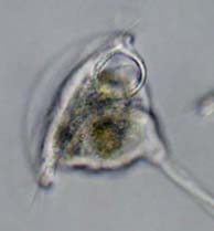
Pyxicola pusilla Wrześniowsky, 1866
similar species - Pyxicola carteri
Ciliophora, Peritrichia.
Found at Patterson Park Lake, Baltimore, MD, USA, the lorica length of sixteen individuals of Pyxicola pusilla measured between ~55 μm and ~80 μm not including the stalk.
This is a loricated species with a short stalk attached to algae. The lorica is widest posteriorly and is crimped anteriorly to allow the operculum to seal the opening when contracted. While feeding, the zooid barely extends beyond the lorica and the operculum almost touches the lorica, unlike the similar species Pyxicola carteri whose peristome extends well beyond the lorica opening.
The macronucleus is a puzzle: there appears to be upper and lower nodes with no apparent connection. Drawings in the literature are disparate and sometimes show a long band-like macronucleus. This was never observed in all sixteen individuals. A somewhat spindle-shaped micronucleus appears below the lower node of the macronucleus. I have not found mention of a micronucleus in the literature.
The infundibulum reaches down to the crimp of the lorica when feeding and a contractile vacuole is just below the peristomial lip. |
 |
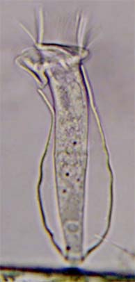
Cothurnia annulata Stokes 1885
Ciliophora, Peritrichia.
Found at Patterson Park Lake, Baltimore, MD, USA, the lorica length of this individual Cothurnia annulata measured ~60 μm including the stalk.
Easy to identify by the ring-like horizontal elevation at about the middle of the zooid. The clear lorica is symmetrically vase-like and attached by a short stalk. There is no valve or operculum. The cell doesn't extend much beyond the lorica opening. When contracted, the cell balls up to a pear-shape within the lorica.
The macronucleus is long and band-like extending above and below the ring-like horizontal elevation; contractile vacuole resides below the peristomial lip and the infundibulum reaches down to about the lorica opening when the cell is fully extended. |
 |
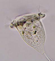
Vorticella astyliformis Foissner, 1981
A species of the Vorticella aquadulcis-complex
Ciliophora, peritrichia.
This is a terrestrial species found in a handful of backyard grass in Baltimore, MD, USA that was soaked in distilled water for a week. The body length of Vorticella astyliformis found measured ~30 μm. The peristomial lip measured ~20 μm.
The distinctive features of V. astyliformis are the roundness of the zooid, a narrow peristomial lip and noticeable striations. The stalk contracts in spiral fashion. Peristomial disc is concave and raised above the peristome at an oblique angle.
A mid-body macronucleus is C-shaped and horizontal to he major axis of the cell. A single large contractile vacuole is directly under the peristome and empties into the infundibulum. The infundibulum is about half the body length. New food vacuoles are round. |
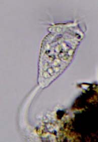 |
Opisthostyla annulata Stokes, 1886
Ciliophora, Peritrichia
Found at Patterson Park Lake, Baltimore, MD, USA, the body length of Opisthostyla annulata found measured between ~20 μm and ~30 μm.
Easy to identify by its hollow stalk which acts like a spring and obvious annulations. Opisthostyla, though solitary, is related to the Epistylididae. The stalk is curved at the attachment and when the zooid is startled, it is thrown backward and recovers in the blink of the eye. The zooid contracts into an oval shape when this happens.
The shape of the zooid is an elongated inverted bell with a peristomial lip and elevated disc. One contractile vacuole is located under the peristomial lip. Macronucleus is "C"-shaped and located in the vicinity of the infundibulum which penetrates the cell about one-third the body length. |
 |
Pseudovorticella chlamydophora (Penard, 1922)
Very similar to Pseudovorticella vestita but with only one contractile vacuole.
Ciliophora, Peritrichia.
Found at Patterson Park Lake, Baltimore, MD, USA, the body length of Pseudovorticella chlamydophora found measured between ~40 μm and ~50 μm.
The most prominent diagnostic feature of P. chlamydophora is the thick clear layer of individual pellicular blisters giving the zooid a quilted appearance and exposing the brick-like silver line system which separates Pseudovorticella species from Vorticella species. The body shape and yellow food vacuoles give the appearance of Vorticella citrina except for the blisters and nucleus shape.
The zooid has a wide peristomial lip. Contraction is complete - the stalk and the cell contract. Contrary to some internet descriptions, the cell is rarely round when it contracts. Instead the cell puckers and contracts into a roughly trapezoidal shape in profile.
The macronucleus is thin and runs along the longitudinal axis of the cell. At times is appears as a weak "C"-shape. One contractile vacuole separates this species from the very similar P. vestita which has two. The infundibulum reaches down almost half the body length.
Pseudocolonies are comprised of individuals. Eats bacteria. |
 |
Pseudovorticella monilata Tatem, 1870
Syn: Vorticella monilata
Ciliophora, Peritrichia
Found at Patterson Park Lake, Baltimore, MD, USA, the body length of Pseudovorticella monilata found measured between ~40μm and ~70 μm long.
Distinctive features of V. monilata are; Pellicular tubercles that look like dots placed along the latitudinal striations, and two contractile vacuoles in the vicinity of the infundibulum which are difficult to detect due to tubercles and food vacuoles. The body is bell-shaped with a wide peristomial lip which sometimes exceeds the body length. A large oral opening leads to an infundibulum that penetrates to about one-third the body length. The macronucleus is "J"-shaped at the bottom, then curves around the peristomial lip a short distance.
Observation of the zooid and stalk contractions yielded surprises: most of the zooids from the same population did not contract and the stalk only contracted one or two coils; zooids that bent to the side but did not contract; zooids whose stalk contracted but the zooid did not; zooids that contract with a coiled stalk. Except of the last iteration, these variations are not typical of Vorticella or Pseudovorticella. |
 |
Vorticella campanula Ehrenberg, 1831
Ciliophora, Peritrichia
Found at Patterson Park Lake, Baltimore, MD, USA, the body length of Vorticella campanula found measures between ~60 μm and ~70 μm long.
Easy to spot due to the dark granules in the cytoplasm, V. campanula has a wide peristomial lip, often exceeding the body length. The body (zooid) shape may vary. Under low magnification the zooids appear almost black.
Though comprised of individuals, pseudocolonies may contain a densely packed group. The stalk contracts in a spiral fashion. Individuals may bend to the point of the peristomial lip almost touching the stalk.
A conspicuous oral opening and undulating membrane lead to an infundibulum that reaches about one-half the zooid length. The long "J"-shaped macronucleus runs more or less longitudinally up the zooid and then follows along the peristomial lip then dips back down into the body. A single contractile vacuole is next to the infundibulum under the peristomial lip and is often difficult to find due to the dark granules.
Uncontaminated freshwater ponds. Eats bacteria. |
 |
Vorticella nebulifera, O.F. Müller, 1786
Syn; Vorticella similis Stokes 1887
A species of the convallaria-complex.
Ciliophora, Peritrichia.
Found at Patterson Park Lake, Baltimore, MD, USA, the body length of Vorticella nebulifera found measure between ~40 μm and ~90 μm long.
Conical bell-shaped body borne upon a stalk that contracts spirally. The peristomial lip is thin and is barely wider than the body. When the zooid contracts it appears as an oval or pear-shape. Sometimes forms pseudocolonies.
"J"-shaped macronucleus continues into the peristomial area. Contractile vacuole is next to the oral opening. |
 |
Vorticella striata Dujardin, 1841
Ciliophora, Peritrichia
Part of the Vorticella aquadulcis-complex
Found at Patterson Park Lake, Baltimore, MD, USA, the body length of Vorticella striata found measure between ~30 μm and ~50 μm long. The zooid may vary in length relative to width.
Usually found as individuals the noticeably striated zooids are slightly constricted under the peristomial lip. Similar to Vorticella microstoma but with a longer body, thicker peristomial lip and a "C"-shaped macronucleus located near the center of the body. A single contractile vacuole is near the oral opening. The stalk varies in length and contracts spirally. Eats bacteria. |
 |
Vorticella microstoma Ehrenberg, 1830
Syn; Vorticella infusionum Dujardin, 1841
Ciliophora, Peritrichia
Found at Patterson Park Lake, Baltimore, MD, USA, the body length of V. microstoma found measure ~20 - 40μm long. The zooid may vary in length and width.
Vase-like body usually wider than the peristomial lip and is noticeably striated. Peristomial disc rises above lip at an oblique angle. The stalk varies in length and contracts in a spiral fashion. The contracted zooid is rounded.
The Macronucleus runs more or less along the longitudinal axis of the cell and is difficult to observe. A single contractile vacuole is found near the oral opening. Eats bacteria. |
 |
Campanella umbellaria Goldfuss, 1820
Ciliophora, Peritrichia.
Found at Patterson Park Lake, Baltimore, MD, USA, specimens of Campanella umbellaria found measure ~130 - 270 μm long. All zooids contain dark granules and may vary in shape.
Each zooid has a broad peristomial lip and bears four or more bands of cilia directing bacteria to the oral opening. The infundibulum reaches deep into the cell, about one-half the body length where new food vacuoles are formed.
Bifurcated non-contractile stalks branch only once bearing two zooids. When a zooid is startled, it folds inward assuming the shape of a ball. Although sessile, some individuals break free from their stalk, develop a posterior trochal band of cilia and swim freely looking for a new place to settle.
A long vermiform nucleus is "C"-shaped and near the peristome. The single water expelling vacuole is located just under the peristomial lip. |
 |
Vorticella citrina (Muller 1773)
Ciliophora, Peritrichia.
A species if the Convallaria complex.
Found at Patterson Park Lake, Baltimore, MD, USA, these specimens of Vorticella citrina measure ~50μm long with a peristome lip ~50-60μm wide.
Vorticella citrina resembles Vorticella campanula but without the dark refractile granules. individual zooids are often tinted yellow.
Vorticella species are difficult to distinguish as the zooids within one species may vary in size and shape due to age, health and reproductive stage. This can be seen within one pseudo-colony. V. citrina does not form colonies as other peritrichs do; instead several individuals may attach to a common substrate forming pseudo-colonies.
The macronucleus is a J-shape that continues into the peristomial lip area. A single small water expelling vacuole is located next to the infundibulum. When startled, the stalk coils spiral fashion within 1/60th second. Feeds on bacteria. |
 |
Epistylis (Ehrenberg, 1830) sp.
Ciliophora, Peritrichia
Found in Patterson Park Lake, Baltimore, MD, USA, these specimens have zooids that ranged between 130μm and 150μm in length.
Similar to Opercularia, but Epistylis has a well-defined peristomial lip. The peristomial disc is clearly offset from the peristomial lip when feeding. The stalk is clear, non-contractile and branched. When startled the zooids contract, especially at the base, in an accordion fashion.
The single water expelling vacuole is a dominant feature just under the peristomial disc. A single macronucleus is somewhat elongate and wraps around the infundibulum at the anterior end. |
 |
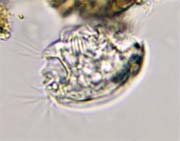
Trichodina (Ehrenberg, 1830) sp.
Ciliophora, Peritrichia.
Found at Patterson Park Lake, Baltimore, MD, USA, these specimens of Trichodina measure ~50μm across and ~20μm thick with the dome. They appeared in the leaking viscous fluid of a crustacean after it was squashed under the cover class.
Free-swimming and in the same sub-class as Vorticella, they are commensal travelers on fish and other aquatic animals while feeding on bacteria. Disease of the host is caused when they are in great enough numbers or enter sensitive areas such as gills or internal organs.
Disc-like in appearance, Trichodina travels swiftly in search of a suitable place to attach. In these specimens the denticular ring is composed of 24 sickle-shaped blades and internal chitinoid hooks. The adhesive disc attaches to a surface by suction.
There are three aboral ciliary rings and one oral ciliary ring. The macronucleus is horseshoe-shaped and circumscribes an almost complete circle under the adhesive disc. A single water expelling vacuole is offset in the center of the disc. The oral apparatus is located between the ends of the macronucleus.
|
 |
Opercularia (Goldfuss, 1820) sp.
Ciliophora, Peritrichia.
These specimens of Opercularia sp. were found in a small ornamental fish pond in Chester, MD, USA. They range in length between 90μm and 100μm. The cell bodies (zooids) are attached to clear, branched, non-contractile and faintly ribbed stalks.
Zooids of these ciliates have a scalloped oral rim lacking a peristomial lip. When the cells are feeding a distinct peristomial disc surrounded by oral ciliature protrudes anteriorly. The oral ciliature guides bacteria and small algae into the infundibulum (also called vestibule). Buccal ciliature within the infundibulum sends the food into a pocket in the cytoplasm which becomes a food vacuole. Adjacent to the infundibulum is a chamber used to eliminate digested vacuoles.
When startled, the stalk does not contract, the peristomial disk recedes into the rim and the zooid moves to the side. The body undergoes minimal contraction except at the base.
A single water expelling vacuole is located next to the lower part of the infundibulum just under the pellicle (cell membrane). The macronucleus is elongate and curved horizontally around the infundibulum. |
 |
Ophryoglena atra (Ehrenberg, 1833)
Ciliophora, Hymenostomatida.
Found in Patterson Park Lake, Baltimore, MD, Ophryoglena atra has an oral apparatus that looks like the number "6" or the capital letter "G." This specimen is 240μm in length, oval and transparent. Ciliation is in dense longitudinal rows. Globules and food vacuoles of various colors fill the cell.
The macronucleus is clear and cigar-shaped located above or near mid-body. The oral apparatus at the anterior end is round with helical membranes that spiral food into the mouth. No water expelling vacuole was reliably observed. |
 |
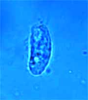
Prorodon teres (Ehrenberg, 1833), Ciliophora, Prorodontidae.
Syn: Holophyra teres
Collected from various ponds around Baltimore, MD, USA, these specimens of Prorodon teres ranged in size from 80μm to 130μm.
Ciliated in longitudinal rows, a dorsal brush (brosse) of three longitudinal rows stands out and extends from cytostome down one-quarter of body length. Trichocysts are numerous around the entire body under the cell membrane. Several long caudal cilia are present trailing the posterior end.
The cytopharynx is supported by numerous trichites that reach deep into the cytoplasm. When the mouth finds an opening into a cyst, the cytopharynx opens wide like a funnel to transfer tissue or nutrients.
Shape depends on diet: starved individuals may be cylindrical or slightly tapered to the posterior; well-fed cells may be ovoid or somewhat rounded, often taking on the color of their last meal. Prorodon teres is histophagous and can be seen here sucking the tissue out of a rotifer cyst.
Macronucleus is round or oval with a clear area in the middle making it look like a fried egg when focus is right. The water expelling vacuole (WEV) is at the posterior end. There may be several small satellite water vacuoles around the WEV. |
 |
Furgasonia (Jankowski, 1964) sp., Ciliophora, Nassulida.
Found at "Lost Lake:" a pond in Patapsco Valley State Park, Maryland, USA. Specimens shown here range between 60μm and 80μm.
Furgasonia is shaped like an elongated oval and longitudinally ciliated with large numbers of perpendicular trichocysts beneath the cell wall (pellicle). The striking array of long trichocysts and bold basket-like cytopharynx makes Furgasonia easy to identify and typical for the genus.
The mouth has visible paroral ciliature followed by an urn-shaped cytopharynx supported by a stiff rod-like basket of trichites. The cytopharyngeal basket may extend through the mouth for ingestion.
The single macronucleus is round and appears below the midline of the cell. A single water expelling vacuole appears at mid-cell near the surface. |
 |
Notohymena (Blatterer & Foissner, 1988) sp.
Ciliophora, Oxytrichidae.
Found in a small stagnant frog pond in Cylburn Arboretum, Baltimore, MD, USA, the specimens of Notohymena shown here range from 140μm - 150μm.
Notohymena has all the attributes of Oxytricha but may be distinguished by the curved peristomial lip in the shape of a hook.
Possible cortical granules were not observed for lack of higher magnification.
Description - Flexible, dorso-ventrally flattened, adoral zone of membranelles (AZM) shaped like a question mark, anterior peristomial lip shaped like a hook, one row each of left and right marginal cirri. The two-nodes of the macronucleus are shaped like elongated ovals, each accompanied by a small dark micronucleus. The water expelling vacuole (WEV) is on the left side at the equator.
18 Ventral cirri: 3 frontal cirri; 1 paroral cirri; 4 frontoventral cirri; 3 post oral cirri; 2 pretransverse cirri and 5 transverse cirri. Caudal cirri are also present. |
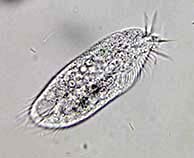 |
Gonostomum affine (Stein, 1859) Sterki, 1878
Ciliophora, Gonostomatidae.
Syn: Oxytricha affinis (Stein, 1859); Gastrostyla affine (Borror, 1972).
The specimens shown are 90μm long and were found in moss from the roof of a backyard shed in Baltimore, Maryland, USA.
The distinctive aspect of Gonostomum is how the adoral zone of membranelles (AZM) follows the left margin of the cell and then arcs abruptly inwards just above the longitudinal midline of the cell unlike species of Oxytricha where the ASM forms a question-mark shape. Another distinction is that the frontoventral cirri remain above the longitudinal midline of the cell. There are no midventral cirri above the pretransverse cirri.
The macronucleus is in two nodes above and below the longitudinal midline. A water expelling vacuole (WEV) near the left margin of the cell is adjacent to the cytopharynx.
There are three frontal cirri, one buccal cirrus, seven frontoventral cirri, two pretransverse cirri and four transverse cirri. Gonostomum has one right and one left marginal rows of cirri. |
 |
Krassniggia (Foissner, 1987) sp.
Ciliophora, Colpodidae.
Found in the moss from the roof of a backyard shed, Baltimore, MD, USA. The carnivorous ciliate has a very large mouth that directs food posteriorly into a pouch before it is channeled into a food vacuole. The size of these specimens ranges from 120μm to 180μm.
Macronucleus mid-body, water expelling vacuole toward the posterior, gaping mouth, uniformly covered with longitudinal and oblique cilia. Mouth cilia is longer than body cilia and creates a swirl current to capture food. |
 |
Green Algae
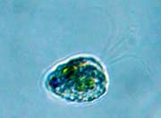
Haematococcus pluvialis (Flotow 1844)
Syn: Haematococcus lacustris
Chlorophyta, Chlorophyceae.
Found in a bird bath in Baltimore, MD, USA, H. pluvialis is common in bird baths, rain pools and shallow freshwater basins.
What looks like a flat disk, is actually a clear ball with the protoplast in the center connected to the cell wall by thin cytoplasmic strands. Watery mucilage occupies the space between protoplast and cell wall.
Two equal flagella (difficult to see) protrude from conical-rounded clear mammilla and cell wall to aid in locomotion. The nucleus is central surrounded by numerous water expelling vacuoles near the surface of the protoplast. A stigma (light sensitive) is behind the anterior end of the protoplast and appears as a black oval.
When H. pluvialis forms a cyst it produces astaxanthin, a blood red pigment that is cultured commercially for cosmetics, dyes and dietary supplements. |
 |
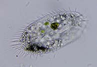
Gastrostyla steinii (Engelmann, 1862)
Ciliophora, Oxytrichidae
Found in a birdbath from Baltimore, MD, USA.
Usually, as depicted in historical drawings, Gastrostyla steinii is elliptical. Though this specimen has all the attributes of G. steinii, it is more or less oval. The overall length is ~150um. The mouth apparatus is about 40-percent of the body length and has two undulating membranes that cross at the midpoint of the mouth.
An unexpected posterior row of four ventral cirri is between the ventral cirri and the right marginal cirri.
Description: The nucleus appears in four nodes; there is no discernable water expelling vacuole for this specimen; two rows of left and right marginal cirri that are continuous at the posterior; three frontal cirri; one buccal cirrus; one paroral cirrus, six frontoventral cirri; two postoral cirri; six ventral cirri; two pre-transverse cirri; and four transverse cirri. Total cirri = 25 not including the right and left rows of marginal cirri or the unexpected posterior row of four cirri. |
 |
Oxytricha longa (Gelei & Szabados, 1950)
Ciliophora, Oxytrichidae.
A large population of these flexible oblong ciliates was found in a birdbath in Baltimore, Maryland, USA. The range in length of the specimens studied was 80-110μm.
Somewhat unusual for Oxytichidae, O. longa has four transverse cirri (where most have five or more) and two long caudal cirri that are offset to the right of the posterior end. The caudal cirri are longer then the overhanging transverse cirri.
Description: O. longa has two rows of marginal cirri that are continuous around the posterior. Two caudal cirri are offset to the right of the cell's posterior and exceed in length the four transverse cirri. The adoral zone of membranelles (AZM) occupies the anterior third of the cell. There are three frontal cirri, a buccal cirrus and four frontoventral cirri. The number of ventral and pre-transverse cirri was undetermined.
A two-node nucleus is located on the left margin of the cell with a water expelling vacuole in between the two nodes. |
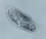 |
Oxytricha fallax (Stein, 1859). Ciliophora, Oxytrichidae.
Found in the scale from the bottom of a birdbath in Baltimore, Maryland, USA, a large population of Oxytricha fallax dominated the fauna.
Members of the Oxytrichidae are difficult to distinguish but clues can help, such as size, number of rows of marginal cirri, frontal cirri, transverse cirri and overall shape.
Oxytricha fallax is a large common Oxytricha; samples in the birdbath population ranged from 130μm to 190μm.
O. fallax in the video has: three frontal cirri; a buccal cirrus; a diagonal row of four frontoventral cirri; three postoral ventral cirri; five transvers cirri and both left and right marginal cirri that tend to lengthen as they approach the posterior.
The cell contains numerous food vacuoles and granules obscuring much of the other internal organelles and making the cell look dark, one water expelling vacuole near the equator on the cell's left side of the body and a macronucleus in two nodes. |
 |
Tachysoma bicirratum (Foissner, Blatterer, Berger & Kohmann, 1991)
Syn: Tachysoma furcata (Kahl, 1932). Ciliophora, Oxytrichidae.
Taxonomic designation of this genera has been under scrutiny.
Tachysoma bicirratum is distinctive by the extraordinarily strong, prominent and long adoral membranelles; long transverse and marginal cirri; and the mouth area that looks like a question mark. This specimen was found in a tiny freshwater frog pond. This individual is ~80μm long.
The posterior-most marginal cirri and the five transverse cirri are extra long giving the appearance of trailing streamers. Strong adoral membranelles are noticeably long throughout the span. The movement of the front membranelles appear as a propeller at times.
Macronucleus is in two nodes located mid-body with a smaller micronucleus in between. The water expelling vacuole is adjacent to the micronucleus on the left outer edge at the equator. |
 |
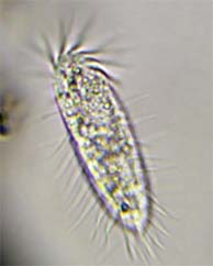
Tachysoma pellionella (O.F. Muller, 1786 Kahl, 1932)
Ciliophora, Oxytrichidae.
Five long transverse cirri and straight marginal sensory bristles make this genera easy to spot, though it can be confused with the genus Oxytricha which bears similar attributes. Tachysoma lacks caudal cirri and the marginal cirri are not continuous at the posterior (as in the genus Oxytricha). Examples in this video are 90μm - 110μm long.
There are three prominent frontal cirri and a single paroral buccal cirrus. The AZM is typical of Oxytricha. Two or more mid-ventral cirri are seen when in focus and five prominent oblique transverse cirri extend well beyond the posterior. The long marginal bristles are straight and stiff. There are two longitudinal rows of ventral-marginal cirri.
The macronucleus consists of two oval nodes separated by a single micronucleus. The marginal water expelling vacuole is adjacent to the micronucleus. |
 |
Stylonychia stylomuscorum (Foissner, Blatterer, Berger & Kohnann 1991)
Syn: Stylonychia muscorum (Kahl, 1932)
Ciliophora, Oxytrichidae.
Found in a small stagnant frog pond, Stylonychia stylomuscorum is one of the smallest of the genera. The individuals found measure 90-110μm. There is a strong AZM that leads to a well-defined paroral membrane. Three stiff caudal cirri extend just beyond the five transverse cirri and the marginal cirri. The left and right marginal cirri get longer toward the posterior but are not continuous. There are four hefty frontal cirri, one paroral cirri, and a diagonal row of three smaller fronto-ventral cirri. Mid-ventral cirri are present. Two macronuclei are in line with the poles and one water expelling vacuole occupies the right side about mid cell. |
 |
Ophryoglena (Ehrenberg, 1831) sp.
Ciliophora, Hymenostomatida.
This round protist has all the earmarks for Ophryoglena: mouth apparatus that looks like the number 6; elongated cigar-shaped nucleus; and a large water expelling vacuole fed by canals. Trichocysts are closely packed beneath the cell membrane below the somatic cilia. The problem is that the cell appears round, unlike the typical Ophryoglena. |
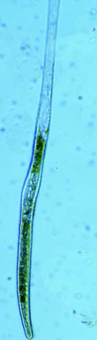 |
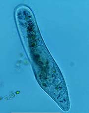
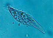
Spirostomum minus (Roux, 1901)
Ciliophora, Heterotrichida.
Spirostomum minus is one of the longest of the genera measuring from about 400μm to 800μm in length, sometimes longer. Individuals are highly contractile and flexible. The peristome is 1/3 the body length and the nucleus appears as a string of pearls. The water expelling vacuole is drawn out up to half the body length. |
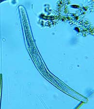 |
Spirostomum teres (Claparede & Lachmann, 1858)
Ciliophora, Heterotrichida.
Widespread in freshwater environments, Spirostomum teres is the smallest of the genera with a slender body length about 150μm to 400μm. The peristome is 1/3 to 1/2 the body length. The centrally located macronucleus is ovoid. The somewhat drawn-out water expelling vacuole is easily seen at the posterior end. |
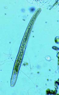 |
Spirostomum intermedium (Kahl, 1932)
Ciliophora, Heterotrichida.
Found in a freshwater pond, this common slender heterotrich is around 400μm to 600μm in length. The peristome is 1/2 the body length and the long macronucleus resembles a chain of beads. The posterior water expelling vacuole is obvious and appears as a colorless feature. A long, sometimes visible water canal feeds the WEV. |
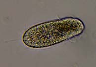 |
Dexiotricha (Stokes, 1885) sp.
Hyemnostomatia, Scuticociliatida.
Medium size hymenostome ciliate 50μm in length and 20μm wide. The cytostome with protruding membranelles occupies the anterior quarter. Heavily ciliated with a caudal cilium ~20μm in length at the rounded posterior. An oblique subapical band of cilia is present behind the truncate cilia-free frontal plate. The spherical nucleus and water expelling vacuole are located mid cell. Feeds on bacteria. |
 |
Diatom
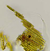
Bacillaria paradoxa (J. F. Gmelin, 1788)
Bacillariophyceae, Bacillariaceae.
The yellow-brown diatom Bacillaria moves around by individual cells sliding along each other on the long axis in stacked colonies giving the appearance of a carpenter's rule. Individual cells contain two chloroplasts with the nucleus in the middle. The colony can extend very far and then retract quickly and change direction in the same manor. The colonies are quite strong and able to drag large clumps of debris. |
 |
Golden algae
Synura spinosa (Korshikov 1929)
Chrysophyceae, Synuraceae.
Found in cold water from a creek in Pine Grove Furnace State Park in PA.
Cells are ~20µm long, colony is ~150µm long.
Individual cells of the Synura spinosa have two long flagella, bristle-like silica scales giving the appearance of cacti and two golden chloroplasts. A colony of Synura spinosa can be either round, or elongate as seen here. The cells, connected at the posterior end, work together as one organism to forage by tumbling around clumps of organic matter. |
 |
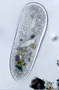
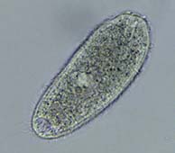
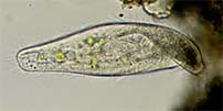
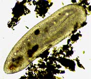
Paramecium caudatum (Ehrenberg, 1838)
Ciliophora, Peniculina.
Paramecia are fairly well known to college grads. They are large cells that be easily seen with a microscope in biology class. The cell is evenly covered with cilia. Between the cilia are rod-like toxic trichocysts which can shoot out like darts to deter predators or immobilize prey. The large faint gray oval macronucleus is often obscured by food vacuoles. There are two Water Expelling Vacuoles (WEV) fed by numerous radiating canals. |
 |
Tillina canalifora (Turner, 1937) Ciliophora, Colpodidae.
Currently, T. canalifora is a synonym of Tillina magna.
Found in the scale of the bottom of a birdbath, Tillina was at first mistaken for a deformed Colpoda.
Tillina cannalifora looks like a Colpoda but has a twisted posterior end making it look somewhat deformed. It has a dark smudge of refractive particles at its posterior end which often obscure the posterior water expelling vacuole (WEV). What look like longitudinal grooves on the surface of the cell are water collecting canals (canalifera) that feed the WEV. The mouth opens to a gullet that spirals down to a food collecting vacuole. A large ellipsoid macronucleus inhabits the mid to anterior region behind the gullet. Several round food vacuoles can be seen moving along the perimeter of the cell. |
 |
Balladyna elongata (Roux, 1901)
Ciliophora, Holostichidae.
The morphology of Balladyna has not been well-studied and its taxonomic designation may have changed. Few species.
Very similar to Tachysoma pellionella but with a large AZM that occupies only 1/4 length of the cell. Two rows of marginal cirri are relatively long and stiff, spaced widely apart. Only one row of ventral cirri. Two nuclei and a single contractile vacuole. Oblique transverse cirri and caudal cirri are present. |
 |
Acineta sp. and Loxophyllum sp.
A suctorian, Acineta sp., tries to ensnare a much larger Loxophyllum sp. The tentacles of the Acineta penetrate the membrane of the ciliate and releases poison to immobilize the cell. The Loxophyllum finally pulls free of the tentacles. |
 |
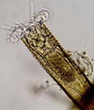
Lacrymaria sp. (Bory de St Vincent, 1824)
Hiding under a testate amoeba.
A novice would have a difficult time trying to identify this combination of two protists. A hidden Lacrymaria is extending its rubbery neck and head from beneath the shell of a testate amoeba. |
 |
Metopus es (O. F. Müller, 1786)
Ciliophora, Heterotrichida.
Common in freshwater and marine environments, Metopus es
forages in the anoxic bottom sediments. This specimen is ~140µm long and ~40µm wide. The frontal lobe is twisted and overhangs the band-like Adoral Zone of Membranelles (AZM). The nucleus is oval shaped and situated adjacent to the AZM just above the equator. The large posterior contractile vacuole is obvious. There is an anterior cluster of dark refractive particles at the tip of the cell. |
 |
Histiobalantium natans (Claparède & Lachmann, 1858)
Ciliophora, Scuticociliatia.
Easily recognizable due to the large mouth, dense cilia and ovoid shape. The mouth has clear membranelles that appear as wisps of silk as they move back and forth. The body cilia are long and bristle-like interspersed with longer groups of cilia. There are two macronuclei and several small contractile vacuoles. |
 |
Leptopharynx (Mermod, 1914) sp.
Ciliophora, Leptopharyngidae.
Found in a sample of grass, this small ciliate (40µm length) can be found foraging on debris feeding on bacteria. Oral apparatus is on the right side about a third down from of the oval shaped cell anterior. Contractile vacuole is adjacent to the oral opening. The spherical macronucleus is centrally located near the contractile vacuole and is difficult to see. |
 |
Metopus spinosus (Kahl, 1927)
Ciliophora, Metopida.
Oddly twisted anteriorly with an anterior lobe overhanging the AZM which spirals down one-third the body length. Anterior tuft of cilia appears on the anterior lobe. Dark granules appear at the front-most part of this lobe. This species has a posterior spine (thus the name "spinosus"). The contractile vacuole is located at the posterior end. M. spinosus has been reported as synonymous with M. attenuatus, M. convexus and M. curvatus. The marine counterpart to this species is M. vestitus, also with a posterior spine (accoring to the National Institute for Environmental Studies, Japan website). |
 |
Anoplophrya (Stein, 1860) sp.
Ciliophora, Astomatia.
Anoplophyra is an Opalinidæ parasite of the guts of annelid worms, especially Oligochaetes. Certain species are found in fish and shrimp. It has also been described in the family Colliniidae. These parasites have no mouth and no apparent means of attachment to their host. |
 |
Frontonia vesiculosa (Da Cunha, 1914)
Ciliophora, Peniculia.
Easily confused with Frontonia leucas, F. vesiculosa has a line of small contractile vacuoles parallel to the cytopharynx. The thin cytopharynx extends from the mouth to the posterior in a straight line. The oval macronucleus appears mid-body but is often obscured by large food such as testate amoebas, diatoms or filamentous cyanobacteria. F. vesiculosa lives in the bottom sediments of lakes and ponds. |
 |
Frontonia Leucas escapes being pinned under a testate Amoeba by creating a hole in its body to isolate the Amoeba. Drama worth watching.
Video has been edited to reduce the time. Total time is about 10-minutes. |
 |
Phialina sp. (Bory de St. Vincent, 1824)
Ciliophora, Litostomatea.
Similar to Lacrymaria, Phialina has an anterior mouth or snout fringed with long cilia and separated from the body by a circular groove. The body is spirally and evenly ciliated. An ovate subcentral nucleus is visible. |
 |
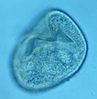
Bursaria truncatella (Müller 1773)
Ciliophora, Colpodea.
A large scoop-like ciliate with large mouth opening that spirals down to the center of the cell. The large ribbon-like nucleus is visible. Bursaria is able to feed on large ciliates, diatoms and flagellates. Brownish color. Able to reach lengths of 1.5mm. |
 |
Mesnilella sp. (Cépède, 1910)
Ciliophora, Hoplitophryidae.
All of the Hoplitophryidae are parasites or commensals of mostly aquatic Oligochaetes and few are to be found outside the intestine of these worms. This ciliate has a clear row of contractile vacuoles and a bluntly truncate and slanted head tapering to a narrow tail. The horn shape of the body is typical as is the mostly backward swimming style.
This video was taken on an old micreoscope and scalebars are unrliable. The organism is about 100µm+ long. |
 |
Cirranter mobilis (Jankowski, 1964)
Ciliophora, Caenomorphidae.
Cirranter mobilis has a very large anterior macronucleus nestled next to a smaller micronucleus. It also has a large posterior contractile vacuole. A band of cilia runs spirally around the body. There are also anterior and posterior tufts of cilia. |
 |
Epalxella antiquorum (Penard, 1922)
Ciliophora, Odontostomatida.
Visually similar to Mylestoma, Epalxella lacks the long posterior cirri. Tufts of long cilia adorn the posterior spines. The mouth area (AZM) has prominent cilia. Posterior spines are similar to Saprodinium. A tooth-like spine is evident above the mouth. Epalxella lives in anaerobic material in bottom pond muck. |
 |
Mylestoma anatinum (Penard, 1922)
Ciliophora, Odontostomatida.
M. anatinum has two visible posterior cirri (fused cilia) and is only about 30µm in length, distinguishing it from the larger genera Saprodinium and Atopodinium. The body is protected by pellicular armor. There is a tooth-like spine above the mouth making it similar to other odontostomes. This ciliate hides under debris while eating bacteria and may be difficult to find. |
 |
Plagiopyla nasuta (Stein, 1860)
Ciliophora, Plagiopylidae.
A noticeable tuft of cilia on the side of the mouth directs bacteria into the cytostome and then into large, plainly visible food vacuoles. Plagiopyla nasuta has been found to contain bacterial symbionts that produce methane. Lives in the muck at the bottom of freshwater ponds. |
 |
Bresslaua vorax (Kahl, 1931)
Ciliophora, Colpodidae.
Similar to other Colpoda species, but Bresslaua has a noticeably gaping mouth. Feeds on other protozoans, algae, fungus, yeast and bacteria, but prefers smaller species of Colpoda. Usually stays near one clump of debris. |
 |
Bothrostoma (Stokes, 1887) sp.
Ciliophora, Metopidae
A large undulating membrane overhangs the cilia around the anteroventral mouth making Bothrostoma unmistakable in appearance. Length = 100µm. A lip, slightly twisted, overhangs the mouth cilia. Posterior contractile vacuole. Caudal cilia are obvious. This ciliate lives in the mud or ooze at the bottom of freshwater ponds and eats organic or decaying matter. |
 |
Saprodinium dentatum (Lauterborn, 1908)
Ciliophora, Odontostomatida.
This ciliate has numerous posterior spines which makes it look loosely like a tiny horseshoe crab and gives it a distinctive appearance. It lives within a carapace. Saprodinium likes to stay close to debris and feed on bacteria. |
 |
Caenomorpha medusula (Perty, 1852)
Ciliophora, Heterotrichida.
Caenomorpha medusula sports a long posterior spine and a medusa-like body which makes it distinctive. A fringe of cilia spirals down to the posterior mouth. Difficult to follow as it swims quickly across the slide or between debris clumps, Letting the sample dry will eventually corral it. Can tolerate low oxygen levels. |
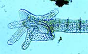 |
Annelid worm
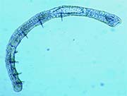
Dero digitata (Müller, 1773)
Oligochaete, Naididae.
David Seamer offered this ID. Dero has finger-like ciliated gills at the posterior end used for gas exchange. The posterior end unfolds like an open hand. The circulatory system carries blood anteriorly on either side of the intestine. Dero Feeds on protozoa, diatoms and other algae, small crustaceans and rotifers. |
 |
Urotricha furcata (Schewiakoff 1892)
Ciliophora, Protostomatea.
This small ciliate usually measures around 30-50µm. Urotricha furcata is easy to spot with its distinctive swimming pattern of leisurely circles or back and forth. A common planktonic ciliate that eats free-swimming bacteria. |
 |
Onychodromus grandis (Stein, 1859)
Hypotrichida, Oxytrichidae.
Easily confused with stylonychia, this genus does not have the three caudal cirri that extend well beyond the body. O. grandis feeds on bacteria and flagellates. Its mouth is broadly triangular. |
 |
Flagellate
Heteronema spirale (Klebs 1893)
Euglenozoa, Peranemidae.
Twisted or spiral body, two flagella, body length 50µm. This flagellate lurks around debris and eats bacteria. |
 |
Didinium nasutum ((Müller, 1773)
Ciliophora, Litostomatea.
Favorite food = Paramecium.
Toxocysts surrounding the cytostome (mouth) immobilize the cilia of Paramecium, or other ciliates when Paramecium are not available. The mouth expands enough to swallow the whole organism. |
 |
Attack of the Dileptids
Two Dileptids relentlessly sting a Chaetogaster (bristle) worm to paralyze it, realize they cannot eat it and move on. It appears that the sting of a Dileptus trichocyst is very painful. Sucking welts are visible from the ciliate trying to consume the worm with suction. Though the video is much shorter, the total time from first sting to abandonment was 10-min. |
 |
Common Bristle Worm
Chaetogaster (Baer, 1827) sp.
Annelida, Naididae.
This genus of Annelid worm has bristles (setae or chaetae) which distinguishes it from other species of Naididae. The bristles are used for crawling along the bottom or on vegetation. Just under the mouth, an array of setae withdraws and protrudes as the worm moves. The mouth can be expanded greatly to accommodate large testate amoebas, rotifers, Euglenas and other protists. The length of this worm varies from 500µm (pers. obs.) to a whopping 25000µm (25mm). |
 |
Homalozoon vermiculare (Stokes 1890)
Ciliophora, Litostomatea.
The anterior mouth of H. vermiculare consists of pharyngeal basket surrounded by poisonous trichocysts which can paralyze prey. The elongated cell has a distinct row of small contractile vacuoles along the length. The most distinctive feature of this protist is the dark and dense parapharyngeal granular mass behind the mouth. It likes to feed on Paramecium, Blepharisma and organic debris. |
 |
Green Algae
Hydrodictyon (Roth, 1797) sp.
Chlorophyceae, Hydrodictyaceae.
The Water Net.
The freshwater Hydrodyctyon cells form an interconnected net with adjacent cells. A distinctive "Y" can be seen where three cells connect. Hydrodictyon reproduces quickly by sexual and asexual means and can cast its net over an entire pond. It is responsible for clogging drains, drainage ditches and disrupting aquatic agriculture such as fisheries. |
 |
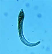
Parasitic Fluke Worm Larva
Platyhelminthes, Trematoda (Rudolphi, 1808) gen. sp.
The cercaria (larval) stage of the fluke worm develops inside a snail. Once released, the free-swimming cercaria finds a host and borrows through the skin. Once an adult is formed, the resulting eggs are excreted in the feces. This fluke was found in a stagnant frog pond with lots of snails. |
 |
Trachelius ovum (Ehrenberg, 1831)
Litostomatea, Tracheliidae.
Trachelius ovum has a short flexible proboscis. Below this protrusion is the cytostome (mouth) that directs food deep into the body. The cytostome can be seen as a round hole like an innertube. According to David Seamer, the two brown balls near the midsection are empty vacuoles. |
 |
Rotifer
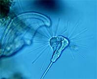
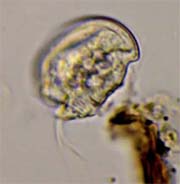
Collotheca (Harring, 1913) sp.
Rotifera, monogonada
Species of the genus Collotheca secrete their own protective tube in which to withdraw when startled. When ready to exit, the infundibulum opens to a gaping yaw. Extra-long cilia along the corona create a wave-like motion sweeping small prey into the mouth. |
 |
Insect larvae
Lake Fly Larvae (also known as midge larvae).
Family: Chironomidae gen. spp.
This family of larvae builds tubes of debris and saliva and uses its body to create a food-bearing current through the tube while protected from predators. |
 |
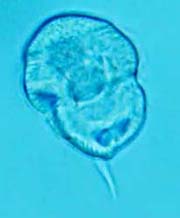
Rotifer
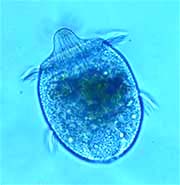
Testudinella patina (Hermann, 1783)
Monogonata, Testudinellidae.
Testudinella patina exhibits the quintessential round body shape of the genus. When the head retracts, so does the tail. The flat loricae allows a nice focal plane of the internal oragans and musculature. When attached to debris, the tail allows the body to rotate 360-degrees.
|
 |
Brachonella (Jankowski, 1964) sp.
Ciliophora, Metopidae.
Brachonella directs bacteria to its mouth with a spiral arrangement of cilia, the Adoral Zone of Membranelles (AZM). The mouth is located posteriorly and dorsally. The anterior black patch is a refractive granule aggregate and the macronucleus is large and ovoid located just below the black aggregate. The contractile vacuole is at the posterior. |
 |
Flagellate
Anthophysa (Bory de Saint-Vincent, 1822) , sp.
Chrysomonada, Chrysophyceae.
Flagellated Colony ~30µm diameter.
If ever there were a tumbleweed of freshwater ponds, the flagellated Anthophysa would fit the bill. Usually found in stalked colonies connected by a sticky colorless substance, colonies break free, usually by sample preparation, and tumble about. Certain cells in a free-swimming colonies can act as a rudder causing the group to change direction. |
 |
Flatworm egg
Gyratrix hermaphroditus (Ehrenberg, 1831)
These stalked egg pods are common in ponds, especially among bottom debris, but can be a mystery as to what they are. A perfectly circumnavigated slit in the pod toward the end away from the stalk provides a "trap door" exit when the worm is developed. |
 |
Flatworm
Gyratrix hermaphroditus (Ehrenberg, 1831)
Platyhelminthes, Polycystididae
Originally labeled as Mesostoma sp., this flatworm belongs to the sub-family Gyratricinae. It has anterior pronounced eyes and a posterior penis stylet. The teardrop greenish eggs are laid with a stalk that attaches to substrate. |
 |
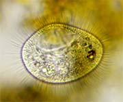
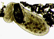
Urocentrum turbo (O.F.Müller, 1786)
Ciliophora, peniculia
Urocenrum spins about its axis and uses cilia to create a swirl current directing suspended bacteria towards the mouth. A tuft of fused cilia forms a "tail." The genera is ubiquitous in freshwater lakes and ponds.
|
 |
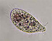
Lembadion (Perty 1849) sp.
Ciliophora, Peniculina
Lembadion has a large oral cavity and can be found rotating gently around the long axis to direct food into its mouth. Feeds on Algae, small ciliates and flagellates. Common in ponds and lakes.
|
 |
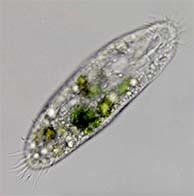
Stentor (Oken, 1815) sp.
Ciliophora, Heterotrichea.
Species of the genus Stentor can assume various shapes as seen here as well as the buccal opening. It can stretch the posterior "foot" to achieve lengths exceeding one or two millimeters making it one of the largest fresh water protozoans in a pond or lake. Collected from the pond in Patterson Park, Baltimore, MD. |
 |
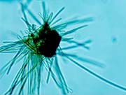
Raphidiophrys (Archer, 1867) elegans (Hertwig & Lesser, 1874)
Heliozoea, Raphidiophryidae.
Raphidiophrys elegans can be found as individuals but are most commonly found in colonies. Look carefully to the cytoplasmic streaming along the "tunnels" connecting the cells. Collected from the pond in Patterson Park, Baltimore, Maryland. |
 |
Flagellate
Collodictyon triciliatum (Carter 1865),
Heterotrophic flagellate,
Chlorophyta, Asteromonadaceae.
Usually described as Algae, this flagellate was originally thought to have three
flagella (thus the species name "triciliatum"). It was later dicovered the flagellate has four. The thin flagella are difficult to see with light microscopy. The shape is variable resulting from occasional bulges in the cell membrane. Research is recommended to discover just how interesting this flagellate is. |
 |
Stylonychia (Ehrenberg, 1830) mytilus,
Ciliophora, Oxytrichidae.
Stylonychia
mytilus is easy to spot for its three long caudal cirri. This ciliate was found amongst the Algae in a lake. Representatives of the genus Stylonychia can be found just about anywhere including dirt, grass, hey, ponds and lakes. |
 |
Stichotricha sp. (Perty, 1849) Ciliophora, Spirofilidae.
Previously identified here as Cohnilembus sp. and corrected by David Seamer, this ciliate prefers to hide its body in debris exposing only the anterior portion to find food. |
 |
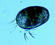
Euplotes sp. (Ehrenberg, 1838) Hypotrichida, Euplotidae
A favorite among protozoa enthusiasts, and somehow satisfying to watch, Euplotes has a deceptively simple design and easy to count cirri or "feet." The Adoral Zone of Membranelles produces a swirl current bringing potential food to its large mouth opening. |
 |
Flagellate
Heteronema sp. (Dujardin, 1841)
Euglenozoa, Peranemaceae.
Heteronema has two flagella: the longer and thicker one pointing forward responsible for swimming and a thinner and shorter trailing flagellum that is usually seen when the flagellate stops or bumps into something. |
 |
Tardigrade
Echiniscus sp. (Schultze, 1840)
Heterotardigrada, Echiniscidae.
The oldest of the family Echiniscidae, the genus Echiniscus might be where we get the term "water bear." It is covered with armor-like plates giving it a dark bear-like appearance. The four claws on each of the eight legs look like those of a bear and it sort of walks like a bear. This species has feelers around the mouth and extra long anterior feelers appearing as thin horns. |
 |
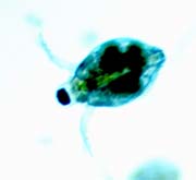
Aspidisca aculeata (Ehrenberg, 1838)
Ciliophora, Aspidiscidae
Aspidisca aculeata sports a thorn on its dorsal side reminiscent of a witches hat. Like all Aspidisca species, it consumes a lot of bacteria.
|
 |
Aspidisca costata, (Dujardin, 1841)
Ciliophora, Aspidiscidae.
Aspidisca is an elegant and beneficial creature living in most fresh water environments. It walks quickly around debris and eats the bacteria it encounters. Aspidisca will often walk upside down on the underside of the cover glass.
|
 |
Blue-green "algae."
Cyanobacteria (Prokaryote) - Formally Blue Green Algae
Blue green “Algae" is not Algae at all. It is the only heterotrophic bacteria that has internal membranes called thylakoids in which photosynthesis takes place, producing oxygen. Cyanobacteria release a significant amount of oxygen into the atmosphere.
Cyanobacteria (blue green "Algae") blooms on summer lakes and stagnant ponds release cyanotoxins which are harmful to animals and humans if ingested.
Cyanobacteria are among the oldest bacteria on earth.
Many forms of Cyanobacteria strands are motile, making it easy for the strands to travel or get tangled into a large mass. |
 |
Seed shrimp
Heterocypris spp.
Crustacea, Ostracoda.
Two species of seed shrimp. Common in lakes and ponds, Heterocypris is bi-valved Crustacean protected within a shell. They multiply quickly and can overcome a fish aquarium if left unchecked. Their sizes range between 150µm (.150mm) to 3mm. |
 |
Lacrymaria Bory de St Vincent, 1824, sp.
An amazing "swan tear" pond microbe.
Lacrymaria sp. can contract or extend its neck many times its own body length. The neck is so flexible, it can quickly probe for food while the body stays relitively still. A must-see protist.
Body length: 100µm
Neck length: up to 700µm
Common in ponds. |
 |
The Water Flea
Scapholeberis sp.
Cladocera, Daphnidae
Crustacean, Brachiopoda.
Also known as Daphnids, these "fleas" are common in lakes and ponds. Larger adults are visible to the eye. The second half of the video shows some of the inner workings and the swim "paddles." |
 |
Flatworm
Catenula (Duges, 1832) sp.
One of the oddest of the flatworms in that during reproduction one zooid develops behind the other forming a chain of offspring. "Catenula" means "short chain" in Latin. |
 |
Coleps sp. - A Voracious Ciliate.
This ciliated scavenger will overcome most cultures if fed properly. Coleps will attack living protozoans and invertebrates and feast on the remains. They can become so numerous, its difficult to view other microbes. |
 |
Tardigrade - A closer look at two common species (Invertebrates).
Macrobiotus sp. and Milnesioum tardigradus are common species of tardigrade found in moss and lichens. This video compares the two using diagnostic features such as buccal apparatus, pharyngeal bulb, placoids and claws. |
 |
Spider Mite (invertebrate)
An arachnid, subclass acarina (mites and ticks) of the family Tetranychidae, this spider mite has unusually long hair-like setae.
Body 250µm = .250 mm |
 |
Amoeba
Platyamoeba placida
From Radiosa to Lobate Form.
Similar to Mayorella sp., Platyamoeba placida is a shape changer. Unlike Mayorella sp., the floating radiosa shape of Platyamoeba maintains a geometric layout of its pseudopodia and the pseudopodia taper to a needle-like point. Similar to other radiosa forms, at least one pseudopoda anchors to the glass slide enabling the amoeba to "walk." |
 |
Amoeba
Mayorella sp.
From Radiosa to Lobate Form.
This Amoeba is a shape changer. The floating version maintains a radiosa form while the locomotive version is lobate. Watch the transformation. While the floating form can be adrift for short periods, it mostly hangs on to either the glass slide or debris with at least one radiating psuedopodia. |
 |
Phacodinium metchnikoff
Cilia Structure and Macronucleus
This recently deceased individual is a perfect specimen for the cilia structure, macronucleus location and food vacuoles of Phacodinium metchnikoffi. The ventral ridge of cilia gives the genus a distinctive appearance. |
 |
Cyclidium sp.
This tiny ciliate has a filtering undulatory membrane that unfurls from its mouth when feeding. While gulping down bacteria, Cyclidium jerks backward from time to time similar to Vorticella. Can be found in water with a high bacteria count.
40X Objective
20-25µm long = .025mm
|
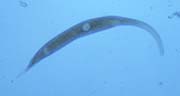 |
Dileptus anser
This ciliated protozoan has a long flailing proboscis (trunk) and a pointed tail. The round mouth, at the base of the trunk, contains poison extrusomes that stun its prey. The mouth can enlarge to accommodate larger prey. D. anser whips its truck around to aid in finding suitable prey. The body and trunk cilia are short and difficult to see, if at all.
10x and 40x objectives
350µm long = .350mm |
 |
Phacodinium sp.
This rare ciliated protozoan, usually found in moss, has a unique cell structure that makes it look like a futuristic UFO. Waves of cilia along its body and around its perimeter help with feeding and locomotion. There is only one genus and several species.
10x Objective
150µm long = .150mm
Found in moss
|
 |
Flagellate
Notoselenus sinuatus
Sometimes mistaken for Petalomonas, spp., but Notosolinus has two flagella. A long flagellum points rigidly forward while mobile and whips around when foraging. A short flagellum trails behind and sometimes appears when the flagellate stops to poke around for bacteria.
40x objective
Length 40µm = .040mm
|
 |
Testate Amoeba
Trinema sp.
Looks like a walking pull tab.
The circular opening of the clear test of this Amoeba makes it look like a pull tab. It walks on hair-like filopodia (pseudopoda) when mobile.
40x objective
Body length ~50µm = .050mm
|
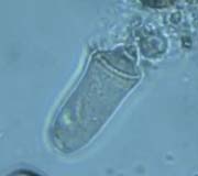 |
Journey of the Vorticella sp. Telotroch (Swarmer) Larva
After asexual binary fission, one of the involved vorticella cells is shorter and develops a broad base unlike its tapered parent. A ring of cilia develops and surrounds the base. The cell eventually breaks free to seek new substrate and start a new colony. The free-swimming cell is called a "telotroch" or "swarmer" and swims with its base forward. Once the teletroch attaches to some debris, the basal cilia are absorbed within minutes as the mouth opening unfurls.
10x, 20x, 100x objectives
Telotroch length ~50µm = .050mm
|
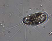 |
Testate Amoeba
Euglypha sp.
This testate amoeba lives in a clear shell (test) covered with excreted plates.While mobile it uses finger-like bendable filopodia for locomotion. The plates around the mouth opening come to a V-shaped points that resemble a circle of teeth.
40x and 100x objectives
Body ~50µm = ~.050mm
|
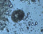 |
Colpoda inflata
Creating swirl current.
This overfed Colpoda inflata uses its cilia to create a current that leads to its mouth. Anything caught up in the swirling water is fair game. But some of the debris in the current is waste from the Colpoda. |
 |
Amoeba
Polychaos Fasciculatum ?
This small Amoeba floats along looking for a place to settle down. The clue to species identification is the numerous short circumferential pseudopodia. The pseudopodia are layered. Another clue is its size.
40x objective
50µm across - .050mm |
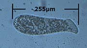 |
Spathidium spathula
This spatulate protist uses its flared mouth to vacuum up bacteria and other protozoans similar to an elephant using its trunk. The lateral viewpoint reveals a dorsoventrally compressed mouth looking somewhat like a spatula.
255µm = .255mm
|
 |
Amoeba
Vahlkampfia sp.
Not your typical amoeba, this one travels by using only one or two pseudopods.
20x objective
45µm long = .045mm
|
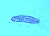 |
Hemicycliostyla sp.
Easily mistaken for a Paramecium, the Hemicycliostyla (Hypotricha, Ciliophora) has several anterior cirri (the appearance of whiskers). This ciliate is very flexible and and uses ventral cilia to "walk" on debris in its search for food.
10x objective
130µm long = .130mm
|
 |
Halteria sp.
Mostly round and skittery, this tiny ciliate can jump many times its body length faster than the blink of the eye. At 200X the first brief sequence is in natural time, the rest are in natural time but the ciliate has been slowed with Ward's Detain.
10x and 40x objectives, blue filter
About 22µm length = .022mm
|
 |
Swarm of bacteria
Look along the margin of the debris to see bacteria swarming. Though these bacteria are mostly harmless and we DO have an immune system, it is a bit unsettling to watch so many in a group. Lots of food for protozoa. When you see something like this, it's time to start another culture.
10x objective
|
 |
Colpoda steinii
Hard to identify. Colpodia steinii has the reniform (kidney) shape of a typical Colpoda but is much smaller. This single cell organism swims very quickly and at times the body structure, caudal cirri, and toothbrush-like rows of cilia in front of the buccal area come into view. Buccal cilia extend perpendicularly beyond the body appearing beard-like at times.
40x objective
20µm in length = .02mm |
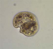 |
Colpoda inflata
40x objective
80µm long = .08mm
|
 |
Colpoda cucullus eliminating Waste, especially at the end of the video. Look for the tiny bubbles and solids floating away.
40x objective
|
 |
Colpoda cucullus
Contractile Vacuole expanding and contracting.
40x objective |
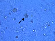 |
This Colpoda undergoes convulsions, contortions and eventually slows down in the process of encysting. Though the progress took about 30-min, removal of sections cut the time to about 6.5-minutes. |
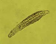 |
Rotifer
Bdelloidea Rotifer (philodina)
4x objective
650µm long = .65mm |
 |
Rotifer
Bdelloidea Rotifer (philodina)
Using Jaws (trophi) and Teeth.
Watch for mastax (pharynx) and trophi (jaw) opening and closing similar to chewing. There are two rod-like teeth on upper and lower jaw. The trophi grind up larger prey and are the only hard parts of the rotifer which are fossilizable.
40x and 10x objectives |
 |
Rotifer
Bdelloidea Rotifer (philodina) Dorsal Feeler
Very short video of the dorsal feeler (sensory antenna) of a Bdelloid rotifer.
10x objective |
 |
Rotifer
Bdelloidea Rotifer (philodina)
With Egg.
Large egg in the abdomen. Notice the trophi and teeth grinding as the rotifer feeds.
10x objective |
 |
Tardigrade
Tardigrade sp.
Also known as water bears or moss piglets.
10x objective
450µm long = .45mm |
 |
Tardigrade
Tardigrade sp.
Watch as the focus racks up and down through this decaying tardigrade: claws,
mouth, pharynx, geometric nerve network, digestive tract, etc.
10x and 40x objectives
|
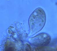 |
Tetrahymena sp.
Feeding on freshly deceased Tardigrade along with bacteria (very tiny).
40x objective
Length of Tetrahymena: 70µm = .07mm
|
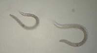 |
Nematoda sp.
C
curling and coiling.
10x objective
Approx. 300µm long = .3mm |
Copyright 2017 - 2018 Steve Cunningham, Baltimore, MD, USA
|